|
||||||||||||||||||||||||||||||||||||||||||
|
||||||||||||||||||||||||||||||||||||||||||
| SisuCHEMA
- Hyperspectral chemical imaging system Go to Hyperspectral Cameras Go to Specim |
 |
Chemical imaging not only creates the knowledg-base for material identification,
it also provides spatial distribution, concentration clustering and other
statistical information.
Speed, simplicity and high performance in near-infrared imaging, are characteristics
of the SisuCHEMA system providing the user with several advantages: high
speed, low heat load from illumination and flexibility with most sample
shapes and sizes. SisuCHEMA is the only chemical imaging technique offering
a direct application path from laboratory to real-time process.
What the SisuCHEMA does
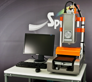
SisuCHEMA is a stand-alone, complete chemical workstation. Samples are
placed into specially-designed sample trays, and by using the ChemaDAQ
data acquisition software, the spectral image is acquired and saved in
seconds. By combining NIR spectroscopy with high-resolution imaging, the
SisuCHEMA provides detailed information on the chemical components, their
quantities and distributions within the sample - invaluable information
for the characterization and quality assurance of advanced materials,
where the functionality of the material is dependent on its chemical and
physical structure.
While the maximum sample size is 200 x 300 x 45 mm, the system can image
samples of 10 mm or smaller at a very high pixel resolution of 30 microns,
and offers flexible settings to coarser resolutions.
SisuCHEMA works like a high speed linescan camera. It acquires and builds
the spectral image of a moving sample line by line, and simultaneously
acquires all wavelengths for each line. This imaging technique is ideal
solution for on-line process monitoring, where samples are in continuous
motion providing a significant advantage to the user. SisuCHEMA employs
pushbroom imaging, acquiring the image one line at a time while scanning
the sample on a moving sample tray. Each line has a 320 pixel field of
view and the variable scanning length allows the user to image longer
samples, or multiple sequential samples, in a single linear scan where
the maximum scanning length is 300 mm.
Easy to configure, simple to use and with a high data capture rate, material-scanning
can be monitored in real time on screen and can provide a direct application
path from the laboratory to real-time process.
Coming pre installed with the UmBio Evince hyperspectral image analysis
software allows users instant application processing, chemical calibrations
and predictions directly within the sisuCHEMA system.
About the camera
SisuCHEMA is based on hyperspectral camera operating in the near-infrared
range with high spectral resolution. Light throughput is 10 to 20 times
higher than similar instruments that implement tunable filters, providing
considerably faster imaging under similar illumination conditions. Requiring
only line illumination allows for a significant reduction of heat load
on the sample. In addition, SPECIM’s unique line illumination technique
optimizes the imaging of various surfaces and textures.
| Applications
|
|||||
| The SisuCHEMA is ideal for pharmaceutical, geological, agricultural applications where high spatial resolution is required and samples are small and on-line process monitoring, where samples are in continuous motion. | |||||
| • Geology | • Tablet analysis | • Blister package inspection | • Blend uniformity | • Food and Dairy | • Forensics |
| • Agricultural Material Screening | • Granule Size and Size Distribution | • Life Science | |||
| Specifications
SisuCHEMA
|
VNIR |
NIR |
SWIR |
| Spectral Range | 400 - 1 000 nm |
900 - 1 700 nm |
1 000 - 2 500 nm |
| Spectral sampling/pixel | 0.78 - 6.27 nm |
4 nm |
6.3 nm |
| Spectral resolution | 2.8 nm |
6 nm |
10 nm |
| Spatial pixels/ line | 1312 |
320 |
|
| Pixel size on sample | 38 - 152 µm |
Scalable from 30
to 600 microns |
|
| Field of view on sample | 50 - 200 mm |
Scalable from 10
to 200 mm |
|
| Maximum sample size | 200 x 300 x 45 mm
(WxLxT) |
||
| Typical scanning time | < 7 s for single
320 x 320 pixel image capture with 256 spectral bands |
||
|
If you like this page, please recommend and share it. |
|||
| More | |||


